Vascular diseases

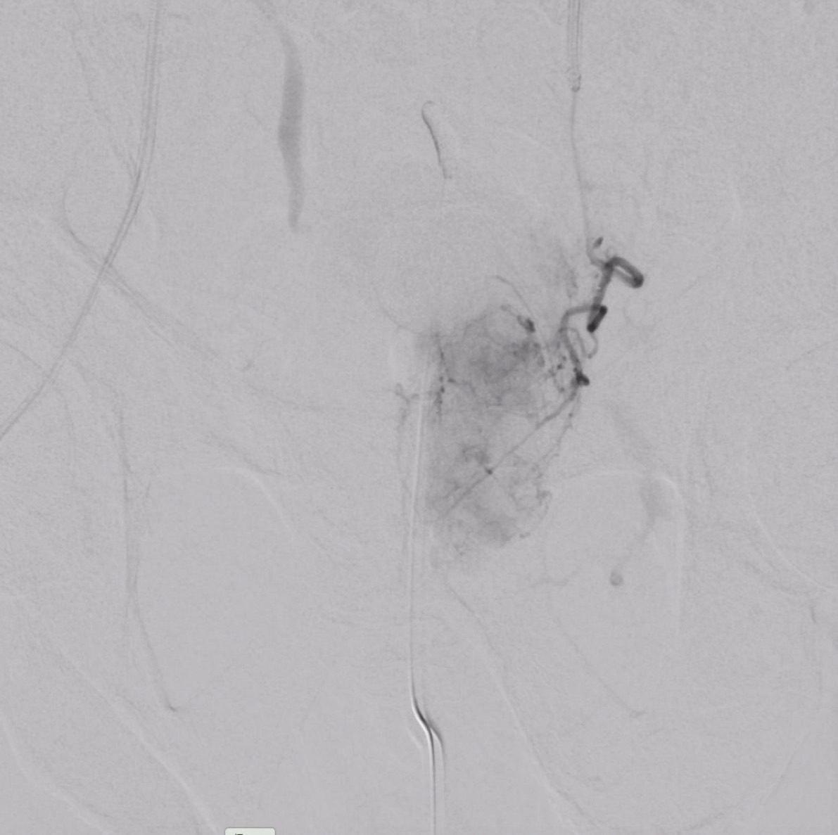
Multiple and sometimes severe and life-threatening vascular diseases can effectively and minimally invasively be treated by interventional radiological methods.

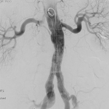
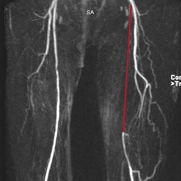
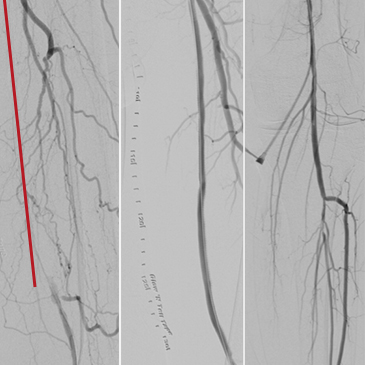
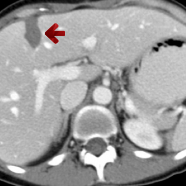
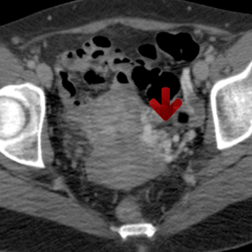
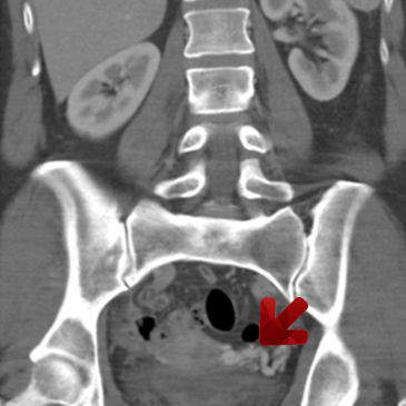
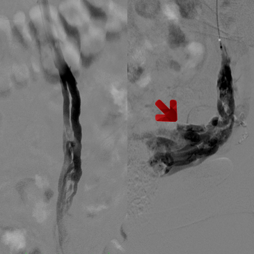
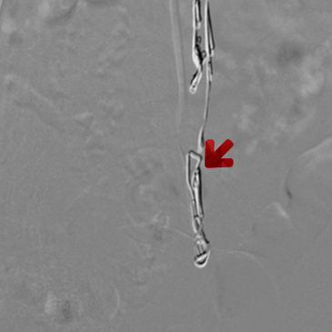
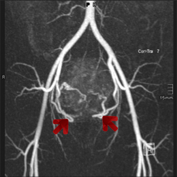
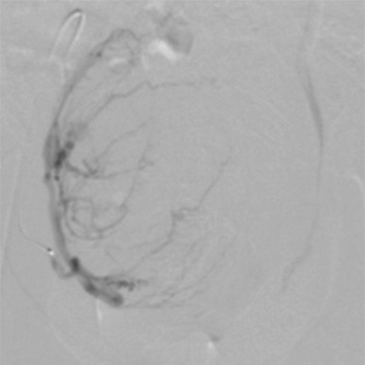
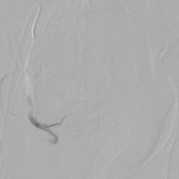
PAOD (=peripheral arterial occlusive disease) as well as occlusive disease in other arterial areas like the visceral, the subclavian, and carotid arteries, are the most common types of vascular diseases that can be treated in a minimally invasive manner.

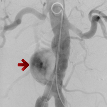
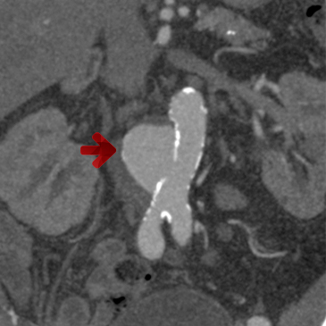
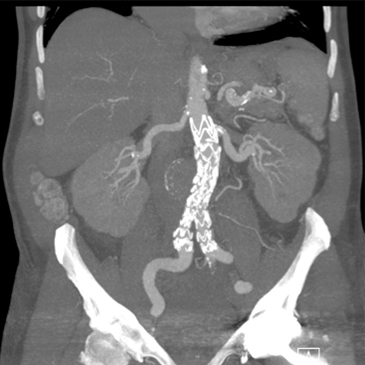
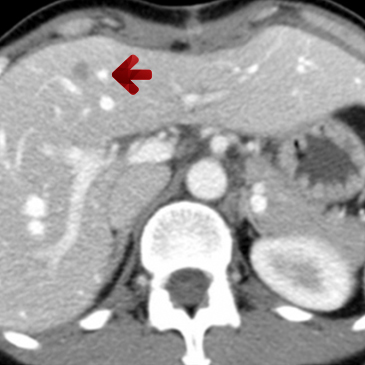
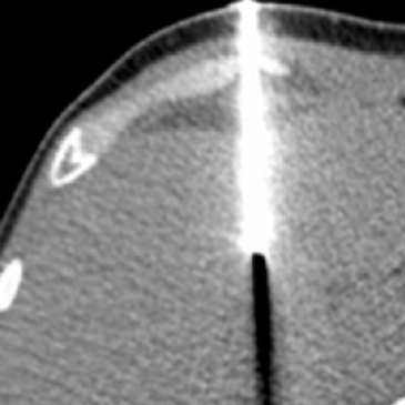
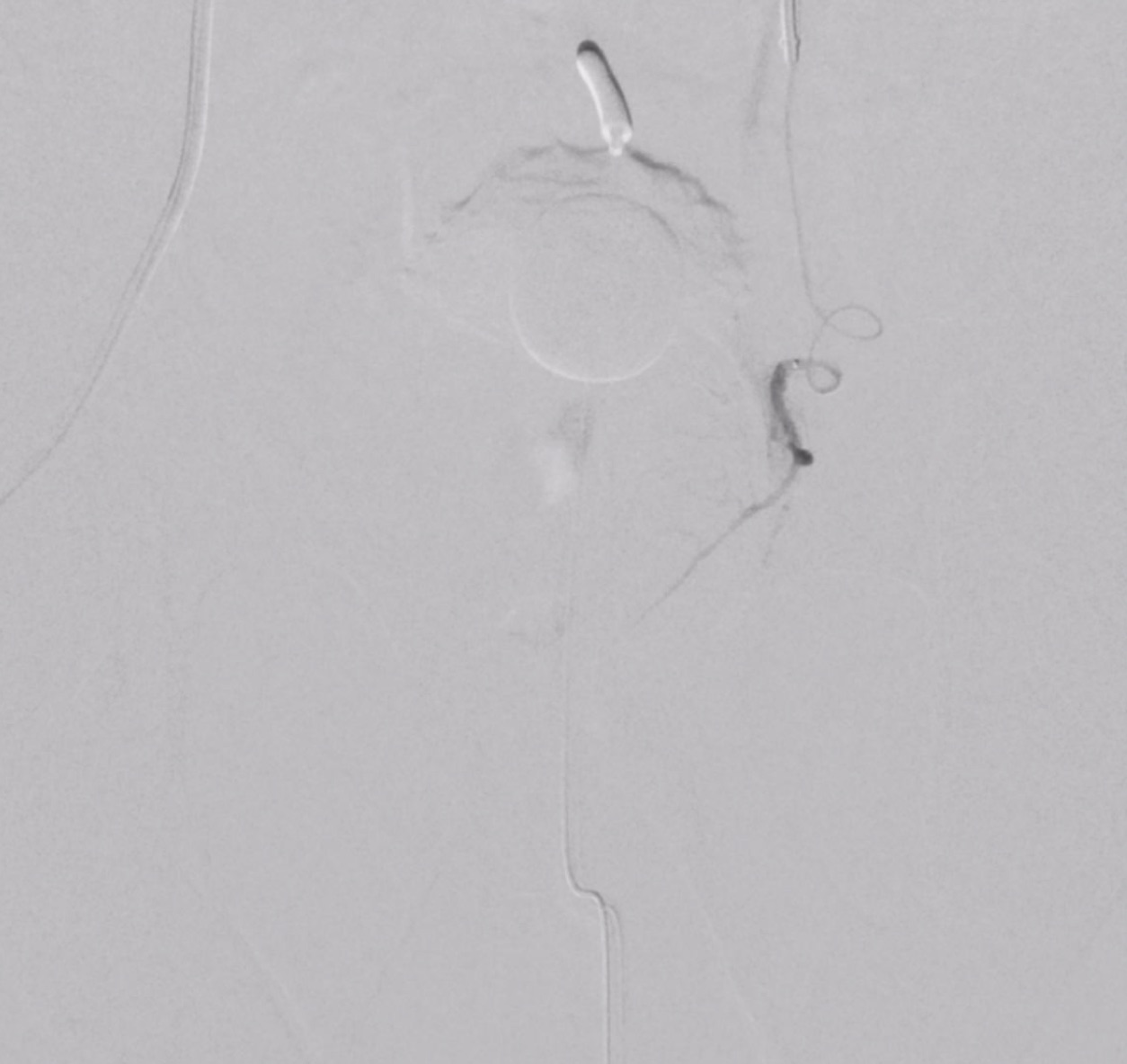
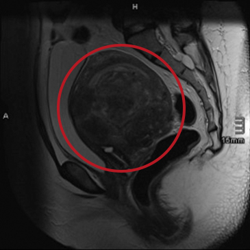
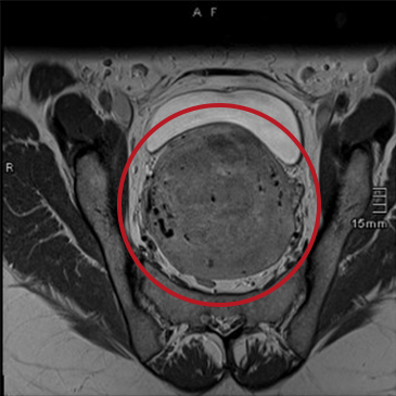
Another very important target for interventional radiology are the diseases of the thoracic and abdominal aorta, and, in particular, the pathological widening of these large vessels (aneurysms). Together with our surgical partners, interventional radiology is also able to treat these diseases highly effectively with minimally invasive methods.
  • Treatment of PAOD patients
    • Balloon Dilatation (PTA = percutaneous transluminal angioplasty), and stent implantation in patients with stenosis or occlusions of the pelvic or peripheral arteries (Image No. 2)
    • Treatment of acute occlusions of the pelvic or peripheral arteries or stents by transluminal thrombectomy /aspiration / lysis
    • Treatment of stenosis of surgical bypasses in PAOD patients
  • Balloon dilatation / stent implantation in the subclavian artery (for example in patients with “subclavian-steal-syndrome”)
  • Endovascular treatment of carotid artery stenosis
  • Endovascular treatment of renal and visceral artery stenosis (celiac trunk, superior mesenteric artery) by balloon dilatation and/or stent implantation
  • Endovascular treatment of stenosis of the abdominal aorta
  • Complete percutaneous treatment of isolated iliac artery aneurysms by stentgraft implantation and/or coil placement
  • Complete percutaneous treatment of visceral and peripheral arteries with stentgraft implantation (covered stent) and/or coil placement
  • Treatment of abdominal and/or thoracic aortic aneurysms with stentgraft implantation (see image No. 1)
  • Treatment of acute dissections of the aorta or other vessels in an acute or chronic setting
  • Mechanical thrombectomy in acute cerebral stroke patients
  • Treatment of venous stenosis with balloon angioplasty and/or stent implantation (for example, pelvic veins or superior vena cava)
  • Recanalization of chronic occlusions of the pelvic veins in patients with chronic post-thrombotic syndrome
  • Embolization of acute arterial bleeding (iatrogenic, traumatic, or spontaneous), for example, in the kidney, the liver, or peripherally, using coils, glue, or particles
  • Treatment of dialysis access stenosis with balloon dilatation
  • Minimally invasive occlusion of a pseudoaneurysm in the common femoral artery after groin puncture
  • Diagnostic angiography, for example, before reconstructive plastic surgery for detailed vessel visualization

Image examples

Endovascular treatment of an abdominal aortic aneurysm
1.1
1.2

Angiography (left image) and CT angiography (right image) of an abdominal aortic aneurysm (red arrow), in danger of rupture because of its size and configuration—immediate therapy for this life-threatening disease is necessary.

1.3
1.4
Angiography (left image) and CT angiography (right image) of the abdominal aorta after minimally invasive, endovascular implantation of a stentgraft. The aneurysm is completely excluded.
Endovascular treatment of an occluded femoral artery
2.1

This patient suffered from a peripheral arterial occlusive disease, with a maximum walking distance of 150m, after which pain began in the region of the left calf. On MR angiography, an occlusion of the left superficial femoral artery can be seen (red line).

2.2

Angiographic image of the vessel occlusion (left image, red line near the occluded femoral artery).

Angiographic image of the femoral artery (right image) after minimally invasive recanalization, dilatation, and stent implantation.

Oncological diseases

Interventional oncology is a relatively young and fast-growing part of interventional radiology. The embolization of tumors, in particular, is performed more and more frequently. It can be done as a single treatment or in combination with surgical therapy, for example, in highly vascularized tumors.
With CT- or ultrasound-guidance, almost all regions of the body can be reached in order to gain a histological specimen, and thus, enable the optimal treatment. The drainage of different fluids from different compartments of the body is also quick and relatively easy with interventional radiological methods. A more and more frequently used method is the CT-guided microwave or radiofrequency ablation of tumors in different regions of the body (for example, liver, lung, kidney).
  • TACE (= trans-arterial chemo-embolization) of the liver in patients with HCC (= hepatocellular carcinoma)
  • Trans-arterial embolization of other primary or secondary liver tumors (for example, cholangiocellular carcinoma, liver metastases in patients with colon cancer, neuro-endocrine tumors, or uveal melanoma, etc.)
  • Embolization of hypervascularized primary tumors or metastases as palliative therapy, or before surgery in order to reduce the intra-operative bleeding risk (for example, in patients with bone metastases from renal cell carcinoma)
  • Radioembolization of the liver (SIRT-therapy) in primary (for example, radio-embolization) or secondary liver tumors (metastases)
  • Percutaneous portal-vein embolization before large liver resections in order to induce hypertrophy of the future liver remnant
  • Implantation of a PICC-catheter (= peripherally inserted central venous catheter) for permanent central-venous access (up to four months)
  • Venous sampling for diagnosis of hormone-producing tumors, for example in the area of the adrenal glands
  • Implantation of a PEG-tube (= percutaneous endoscopic gastrostomy)
  • PTBD (= percutaneous trans-hepatic biliary drainage) – drainage of the bile ducts inward to the duodenum or outward, or stent-placement in the bile ducts in patients with malignant stenosis
  • CT- or ultrasound-guided diagnostic puncture of tumors
  • CT- or ultrasound-guided drainage of pleural effusion or abdominal ascites
  • CT- or ultrasound-guided drainage of fluid collections
  • CT-guided radiofrequency or microwave ablation or primary or secondary tumors of the liver or other organs (see image No. 3)

Image examples

Minimally invasive treatment of a liver metastasis
3.1

Liver metastasis of a colon carcinoma – imaging with computed tomography (red arrow).

3.2

CT-guided radiofrequency ablation of the metastasis – the image shows the metallic probe that is used to “burn” the metastasis.

3.1
CT examination one year after radiofrequency ablation – the necrotic area (arrow) is well visible, there is no vital tumor tissue. No more metastases can be seen in the rest of the liver.

Different benign diseases

Multiple benign diseases can be treated by interventional radiology methods. The embolization of uterine fibroids in patients with symptomatic uterine fibroids is highly effective and minimally invasive, as is prostate embolization in patients with symptomatic benign prostate hyperplasia.
The embolization of the spermatic vein in male patients with a symptomatic varicocele, as well as the ovarian vein in female patients with a pelvic congestion syndrome are also highly effective treatment methods.
  • Embolization of uterine fibroids in symptomatic female pateints (for example, pain, bleeding, etc., see image example No. 5)
  • Embolization of the prostate gland in patients with benign prostatic hyperplasia
  • Embolization of the spermatic vein in patients with a symptomatic varicocele
  • Embolization of the ovarian vein in female patients with pelvic congestion syndrome (see image example No. 4)
  • Treatment of vascular malformations (venous, lymphatic, arteriovenous, etc.) using different techniques such as percutaneous sclerosis, embolization, etc.
  • Implantation and/or removal of vena cava filters for pulmonary embolism prevention
  • TIPSS procedure (= transjugular intrahepatic portosystemic shunt) in pateints with symptomatic portal hypertension
  • PTBD (= percutaneous transhepatic biliary drainage)  – drainage of the bile ducts inward to the duodenum or outward, or stent-placement
  • Minimally invasive removal of foreign bodies (for example, lost catheter fragments, wire fragments, or port-a-cath parts, etc.)

Image examples

Prostate Embolisation in a patient with Benign Prostate Hyperplasia (BPH)
pae-vor-embo-ct-axial
pae-dyna-ct-vorher
Computed Tomography Scan of an enlarged prostate in a 70 y/o male patient presenting with severe symptoms of BPH (=Benign Prostate Hyperplasia) with nycturia, weak urinary stream and urine dropping/leaking.

As treatment of the BPH a prostate artery embolization is planned. The goal of this procedure is to reduce the arterial perfusion of the prostate in order to generate a shrinking of the prostate.

The Prostate Artery Embolization is performed under local anesthesia via the right or left  common femoral artery, the procedure is not painful at all. The present image shows a “Dyna-CT” visualization of the left prostate lobe during the embolization – using this technique the arterial perfusion of the prostate can be visualized and as a consequence the supplying artery can be occluded using small particles.
pae-links-vor-embo

Angiographic visualization of the left prostate lobe using conventional angiography. The micro catheter is already in place and the embolization procedure (=very slow injection of small particles) can be started.

pae-links-nach-embo

Final angiography of the left prostate lobe – it can not be seen anymore, the embolization procedure was successful. The same technique is used on the right side. The patient mentioned above left the hospital the day after the procedure and was asymptomatic a few days later.

Sclerosis of the ovarian vein in a patient with pelvic congestion syndrome
4.1
4.2
Computed tomography of a female patient with pelvic congestion syndrome and unclear pelvic pain, as well as enlarged venous vessels around the uterus (red arrows in the right and left image) and heavily enlarged ovarian vein.
4.3

Angiographic imaging of the ovarian vein after contrast media application in the left renal vein – extensive back-flow in the pelvic veins, visualization of the enlarged peri-uterine veins (red arrow).

4.4

Morphological result after sclerosis and embolization of the ovarian vein. Consecutively after contrast media injection in the left renal vein the ovarian vein and the veins around the uterus are not visible anymore.. Well visible are the metallic coils in the ovarian vein. The patient showed no more pelvic-congestion syndrome after the treatment.

Embolization of uterine fibroids
5.1
5.2
5.3

Magnetic resonance tomography of a uterus with large uterine fibroids (red circles). In the right image, an MR angiography shows the vessel supply of the uterine fibroids via both uterine arteries (red arrows).

4
5

Angiography of the uterine artery on the right side before (left) and after (right) embolization with bland particles.

6
7
Magnetic resonance imaging two weeks after the embolization – the total volume of the uterus has shrunk, the large central uterine fibroid is no longer perfused (necrotic, dark). Two weeks after the treatment, the patient had no more symptoms.

Pain management

Interventional radiological pain management includes modern, highly effective, and safe imaging methods, such as CT and ultrasound.
It is effective in different parts of the body, including the spine in patients with a narrowing of a neuroforamen, osteoarthritis of the intervertebral joints, or symptomatic intervertebral disc extrusion.
  • CT-guided pain management in patients with intervertebral disc herniation (cervical, thoracic, and lumbar spine)
  • CT-guided pain management in facet joint arthritis (small joints in the lumbar spine)
  • Radiofrequency ablation of osteoid-osteomas
  • Vertebroplasty in patients with symptomatic osteoporotic or malignant fractures of vertebral bodies (in cooperation with Ivan Dobrocky, MD, PhD, MSc)